
Figure 4. MSCs suppress ocular inflammation through expansion of myeloid progenitor cells in a CD200-dependent manner.
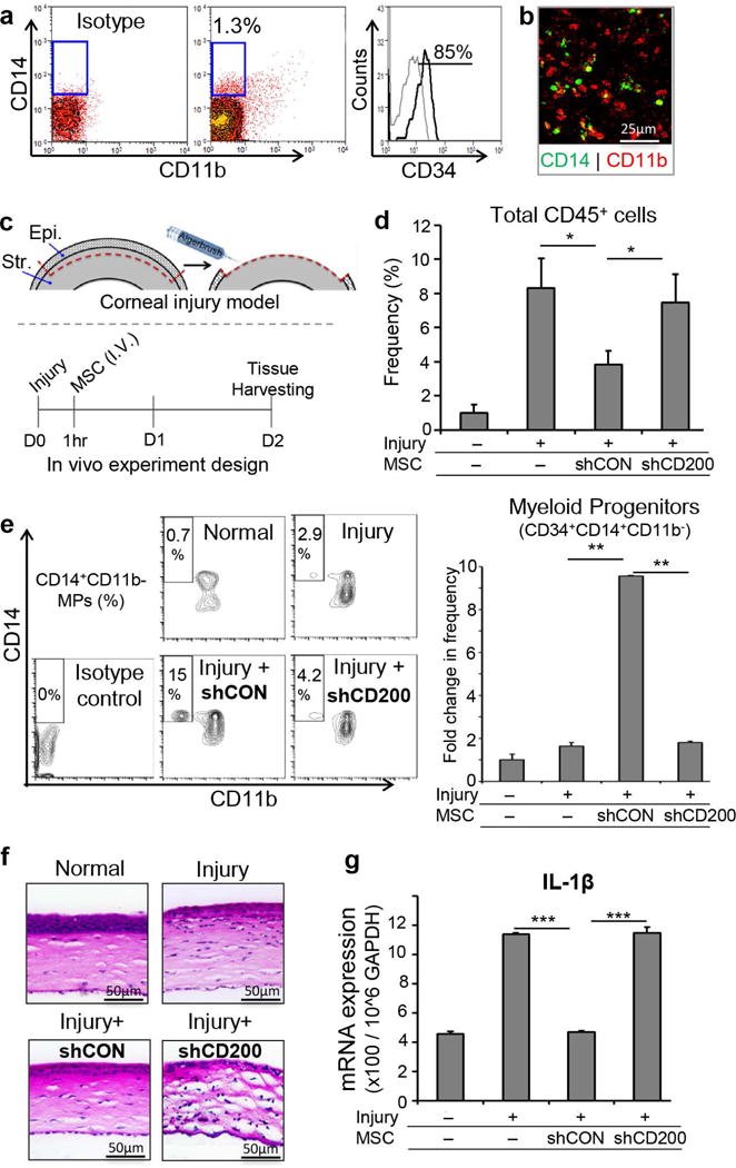
A. Representative flow cytometric plots demonstrating CD34+CD14+CD11b− myeloid progenitor in the cornea. B. Confocal microscopy image (×20 magnification) of corneal whole mount confirming the presence of CD14+ CD11b− cells in the peripheral corneal stroma (Green: CD14, Red: CD11b). C. Schematic representation of sterile injury induction in mouse and experiment timeline. Corneal epithelium and anterior stroma are mechanically removed using Algerbrush II. D. Bar chart demonstrating the frequencies of infiltrating corneal CD45+ cells in naïve mouse, injured mice without systemic MSC treatment, control-shRNA (shCON) MSC-treated and CD200-shRNA (shCD200) MSC-treated mice. E. Representative flow cytometric plots and bar chart demonstrating the frequencies of myeloid progenitors in naïve cornea, injured cornea, injured cornea with IV administration of control-shRNA-treated MSCs, and injured cornea with IV administration of CD200-shRNA-treated MSCs 48 hours after injury induction. F. H &E staining of corneal cross-sections (×20) from naïve, untreated, control-shRNA MSC-treated and CD200-shRNA MSC-treated mice demonstrating epithelial and stromal layers and inflammatory cell infiltration. G. Real-time PCR analysis of relative expression of IL-1β mRNA in naïve mice, injured mice without systemic MSC treatment, control-shRNA MSC-treated and CD200-shRNA MSC-treated mice. Results are representative of 2 independent experiments. Each group consisted of 4–5 animals in each experiment. P values are calculated using student’s t-test and data is represented as mean ± SEM. * p< 0.05, ** p< 0.01, *** p< 0.001