Figure 1.
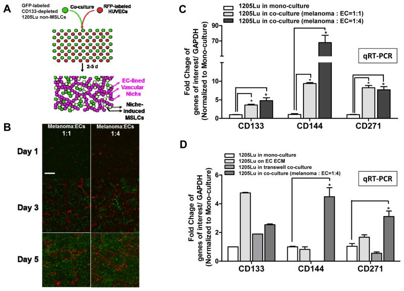
Two dimensional (2D) melanoma-EC co-culture model recapitulates MSLC niche in vitro. A. Schematic representation of the 2D MSLC niche model in vitro. GFP-labeled CD133− non-MSLCs were co-cultured with RFP-labeled human umbilical vein endothelial cells (HUVECs) at 1:1 or 1:4 ratios for 5 days. B. In this model, ECs aligned to form branching tubular networks, reminiscent of the vascular niche in vivo (Magnification, ×100; scale bar, 200 μm). Co-cultured melanoma cells were then segregated from ECs by flow cytometry. C. MSLC (e.g., CD133 and CD271) and VM (e.g., CD144) markers were up-regulated in co-cultured melanoma cells compared to their mono-culture counter parts using qRT-PCR, simulating “dynamic stemness” and VM morphogenesis in vitro. D. Such a niche-inducing phenomenon was most pronounced when melanoma-EC contact is permitted, while EC extracellular matrix (ECM) or soluble factors in transwell cultures alone exhibited limited/partial effects. *, P < 0.05.

