Figure 1.

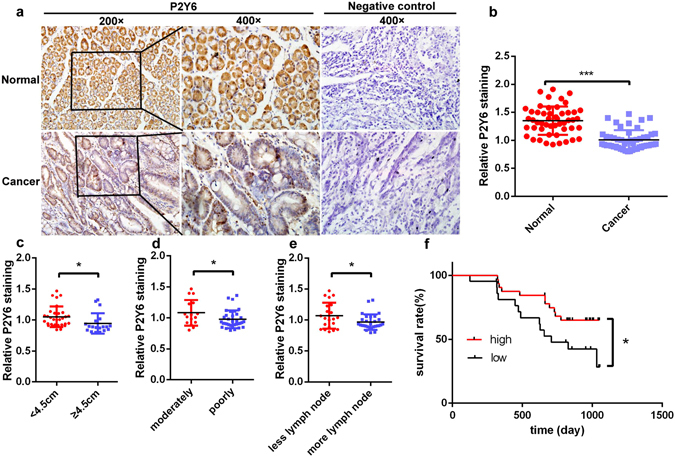
The expression of P2Y6 receptors in human gastric cancer cells and its association with cancer progression. (a) Immunohistochemical staining of P2Y6 receptor expression in human GC tissues and adjacent normal tissues. (b) Summary data showing the reduced expression of P2Y6 receptors in GC tissues compared to adjacent normal tissues. (c–e) The expression levels of P2Y6 receptors in GC samples from the patients with different tumor sizes, differentiation and metastasis to lymph nodes. (f) Kaplan-Meier analysis of different expression levels of P2Y6 receptors with the survival of GC patients. *p < 0.05, ***p < 0.001, n = 50. Image-Pro Plus 6.0 was used to analysis the P2Y6 receptor expression of immunohistochemical staining.
