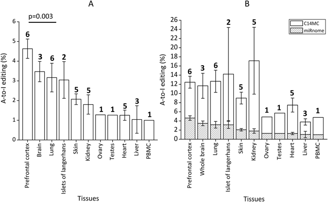
Figure 1.

Distribution of A-to-I editing in different healthy human tissues. (A) A-to-I editing in mature miRNAs is the highest in Prefrontal cortex (two-tailed t-test; p = 0.003 with respect to lung samples). Percentage A-to-I editing was calculated by dividing the number of edited miRNAs by the total number of miRNAs expressed with a read count greater than equal to 10. The numbers above the bars represent the number of different individuals analysed. (B) C14 miRNA cluster show enriched A-to-I editing. The fraction of edited miRNAs from C14 was significantly higher compared to the miRnome average in all tissues analyzed (p < 0.008), the tissues have been arranged according to descending order of miRnome-wide editing.
