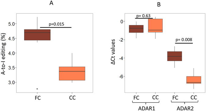
Figure 3.

Neuron rich frontal cortex (FC) showed higher A-to-I editing in mature miRNAs than the corresponding corpus callosum (CC) of the same individuals. (A) FC showed higher A-to-I editing than CC of the same individuals (two-tailed t-test; p = 0.015). (B) Real-time PCR was done and delta Ct was plotted, ADAR2 (and not ADAR1, two-tailed t-test; p = 0.63) showed significant upregulation in FC compared to CC samples (two-tailed t-test; p = 0.008). B2M was used to normalize expression in all samples.
