Figure 6.
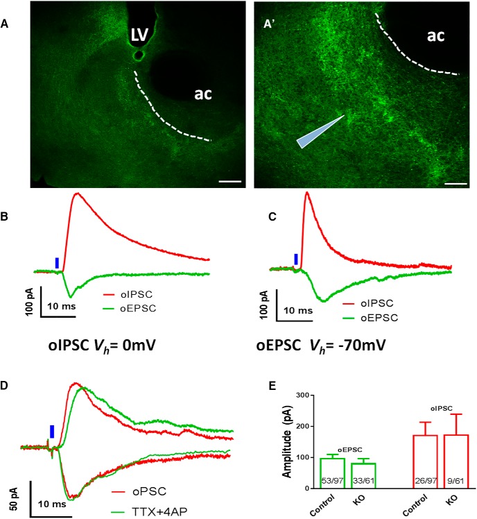
Effects of VMAT2 deletion in VTA LepR neurons on synaptic neurotransmitter release to the accumbens. Recordings of oEPCSc and oIPSCs were made in accumbens neurons elicited by blue laser stimulation of ChR2-expressing fibers from VTA LepR neurons in brain slices. Representative pictures showing ChR2-expressing fibers in the accumbens in low (A) and high magnification (A’). Representative recording traces of light evoked oEPSCs and oIPSCs from brain slices of control (B) and LIC::Vmat2fl°x/fl°x (KO) mice (C). D, Representative traces showing responses of light-evoked oEPSCs and oIPSCs to TTX/4-AP in controls. E, Mean amplitudes of light evoked oIPSC (red) and oEPSC (green) in controls and LIC::Vmat2fl°x/fl°x mice; numbers in the bar showed frequency of successful detection of the indicated currents in recorded neurons. Data were presented as mean ± SEM; ac, anterior commissure; LV, lateral ventricle. Scale bars, 100 μm (A) and 50 μm (A’).

