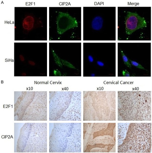
Figure 4.

Sub-cellular co-expression of E2F1 and CIP2A in vivo. A. Representative immunofluorescence staining of CIP2A (green) and E2F1 (red) protein in HeLa and SiHa cells by confocal laser scanning microscopy. The nucleus was stained blue by DAPI. B. Immunohistochemistry staining of E2F1 and CIP2A protein in paraffin-embedded cervical tissue. Positive staining for E2F1 was defined as brown stain in the nucleus and for CIP2A in the cytoplasm.
