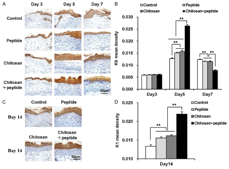
Figure 4.

Peptide-modified chitosan hydrogels promote the proliferation and differentiation of keratinocytes in skin wounds. A: Immunohistochemical staining to show K6 expression in keratinocytes in the control, peptide, chitosan hydrogel, and peptide + chitosan hydrogel groups 3, 5, and 7 days after the initial treatments (scale bar: 50 μm). B: K6 expression in the control, peptide, chitosan hydrogel, and peptide + chitosan hydrogel groups on days 3, 5, and 7 after the initial treatments (n = 3). C: Immunohistochemical staining of K1 expression in keratinocytes in the control, peptide, chitosan hydrogel, and peptide + chitosan hydrogel groups on day 14 after the initial treatments (scale bar: 50 μm). D: K1 expression in the control, peptide, chitosan hydrogel, and peptide + chitosan hydrogel groups on day 14 after the initial treatments (n = 3).
