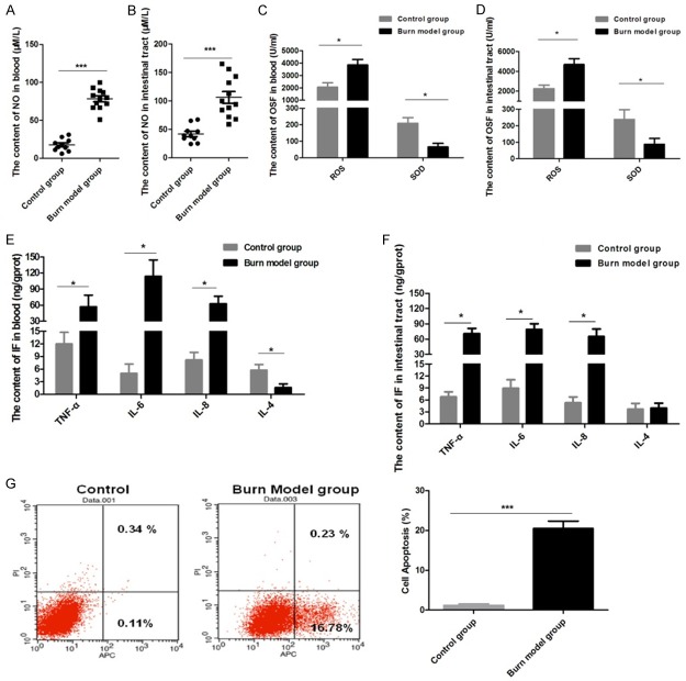
Figure 2.
In blood and intestinal tissue samples, the content of nitric oxide (NO), oxidative stress factors, and inflammatory factors (IF) were significantly increased and the apoptosis rate of the intestinal epithelial cells was increased in the burn model group: A: NO content detected in blood. B: NO content detected in intestinal tissue. C: OSFs were detected in blood. D: OSFs were detected in intestinal tissue. E: IFs were detected in blood. F: IFs were detected in intestinal tissue. G: The apoptosis ratio of intestinal epithelial cells was determined by flow cytometry. Data are presented as the mean ± standard error of measurement (SEM) (n = 3) with *P < 0.05 or ***P < 0.001, analyzed by two-way ANOVA.

