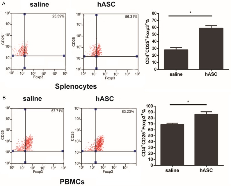
Figure 6.

Induction of Tregs by hASCs in mice with CIA. Cells were activated with PMA and BFA for 4 h in the experimental groups. The splenocytes and PBMCs were stained with CD4, CD25, and Foxp3. A: Expression of Tregs was significantly increased in the spleen as analyzed by flow cytometry. B: Percentages of CD4+CD25+Foxp3+ cells in the peripheral blood in the hASC-treated group were higher than those in the saline control group. *P<0.05. Data are expressed as Means ± SD.
