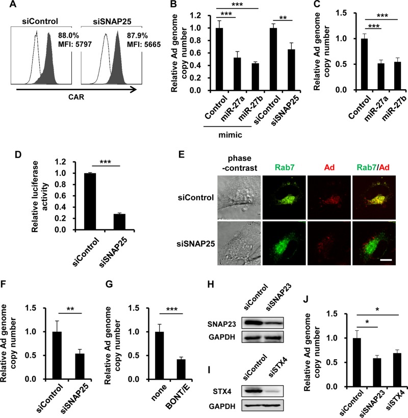
FIG 4.
Inhibition of Ad entry by knockdown of SNAP25. (A) HeLa cells were transfected with siSNAP25 at 50 nM. After 48 h of incubation, the cells were collected and analyzed by flow cytometry for the expression of coxsackievirus-adenovirus receptor (CAR). Open histograms represent isotype control staining. Filled histograms represent specific staining with an anti-CAR antibody. MFI, mean fluorescence intensity. (B and C) HeLa cells (B) and HUVECs (C) were transfected with miR-27a/b mimics at 20 nM or with siSNAP25 at 50 nM, followed by infection with WT-Ad at 100 VP/cell. After 3 h of incubation, the copy numbers of WT-Ad genomic DNA in the cells were determined by quantitative PCR analysis. (D) HeLa cells were transfected with siSNAP25 at 50 nM, followed by transduction with Ad-Luc at 100 VP/cell. After 12 h of incubation, luciferase activities in the cells were determined. (E) HeLa cells were cotransfected with a plasmid expressing a fusion protein of GFP and Rab7 (pGFP-Rab7) and with siSNAP25 at 50 nM, followed by infection with the Cy3-labeled Ad at 100 VP/cell. After 1 h of incubation, phase-contrast, GFP fluorescence (Rab7), and Cy3 fluorescence (Ad) photomicrographs of the cells were obtained. Representative images from multiple experiments are shown. Bar, 10 μm. (F) HeLa cells were transfected with siSNAP25 at 50 nM, followed by infection with a ts1 mutant Ad at 100 VP/cell. After 3 h of incubation, the copy numbers of WT-Ad genomic DNA in the cells were determined. (G) HeLa cells were pretreated with recombinant BONT-E LC at 50 nM for 12 h, followed by infection with WT-Ad at 100 VP/cell. After 24 h of incubation, the copy numbers of WT-Ad genomic DNA in the cells were determined. (H and I) HeLa cells were transfected with the indicated siRNAs at 50 nM. After 48 h of incubation, SNAP23 (H) and STX4 (I) protein levels were determined by Western blotting. (J) HeLa cells were transfected with the indicated siRNAs at 50 nM, followed by infection with WT-Ad at 100 VP/cell. After 24 h of incubation, the copy numbers of WT-Ad genomic DNA in the cells were determined. The data are expressed as means and SD (n = 3 or 4). *, P < 0.05; **, P < 0.01; ***, P < 0.001.

