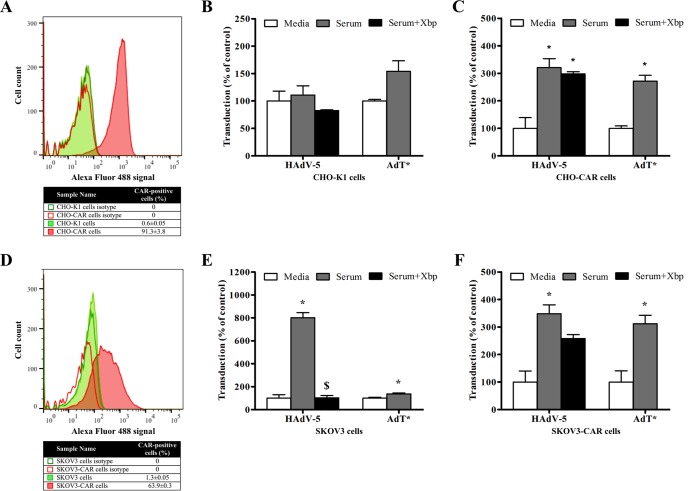
FIG 3.
Adenoviral transduction in the presence of immunocompromised mouse serum in different cell lines engineered to express CAR. (A and D) CAR expression levels on cell plasma membrane were tested by flow cytometry in CHO-K1 and CHO-CAR cells (A) and SKOV3 and SKOV3-CAR cells (D). CAR-positive cells are expressed as a percentage of the parental population and the mean of technical triplicates ± SEM. Representative data are shown. (B, C, E, and F) HAdV-5 or AdT* (FX binding-deficient) (2 × 10¹⁰ vp/ml) was incubated for 30 min at 37°C with serum-free (SF) media or 90% Rag 2−/− serum in the presence or absence of X-bp (40 μg/ml). Adenovirus suspensions were added to CHO-K1 (B), CHO-CAR (C), SKOV3 (E), or SKOV3-CAR (F) cells (MOI, 1,000 vp/cell) and incubated at 37°C for 2 h. Then, the medium was replaced with media containing 2% FCS, and the cells were incubated for an additional 20 h. β-Galactosidase expression levels were quantified as relative light units (RLU) and normalized to the total milligrams of protein. The data represent pooled values from three (B, C, and F) or four (E) independent experiments with four replicates per condition. Values are shown as a percentage of the SF-medium-alone condition and expressed as the mean of the normalized values per experiment plus SEM. Transduction values (RLU/mg of total protein) for HAdV-5 in the presence of SF media from a representative independent experiment are indicated below to add clarity on the magnitude of transduction levels: 4.8 × 10⁴ (CHO-K1 cells), 3.8 × 10⁶ (CHO-CAR cells). Repeated-measures ANOVA and posthoc Tukey's range test were applied. *, P < 0.05 versus matched controls; $, P < 0.05 versus serum.

