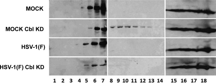
FIG 5.

Changes in the membrane distribution of Nectin-1 in Cbl-depleted cells. HEp-2 cells and their Cbl-depleted derivatives were either mock infected or exposed to HSV-1(F) (10 PFU/cell). The cells were harvested at 8 h postinfection, and detergent-insoluble membranes were adjusted to 1.5 M sucrose and overlaid with 5 ml of 1.2 M sucrose, followed by a 2-ml layer of 0.15 M sucrose. Following ultracentrifugation as detailed in the Materials and Methods section, fractions (500 μl) were collected from the top to the bottom of the gradient. Equal volumes from each fraction were used for protein analysis in 9% denaturing polyacrylamide gels. Immunoblotting was done with the Nectin-1 antibody.
