FIG 1.
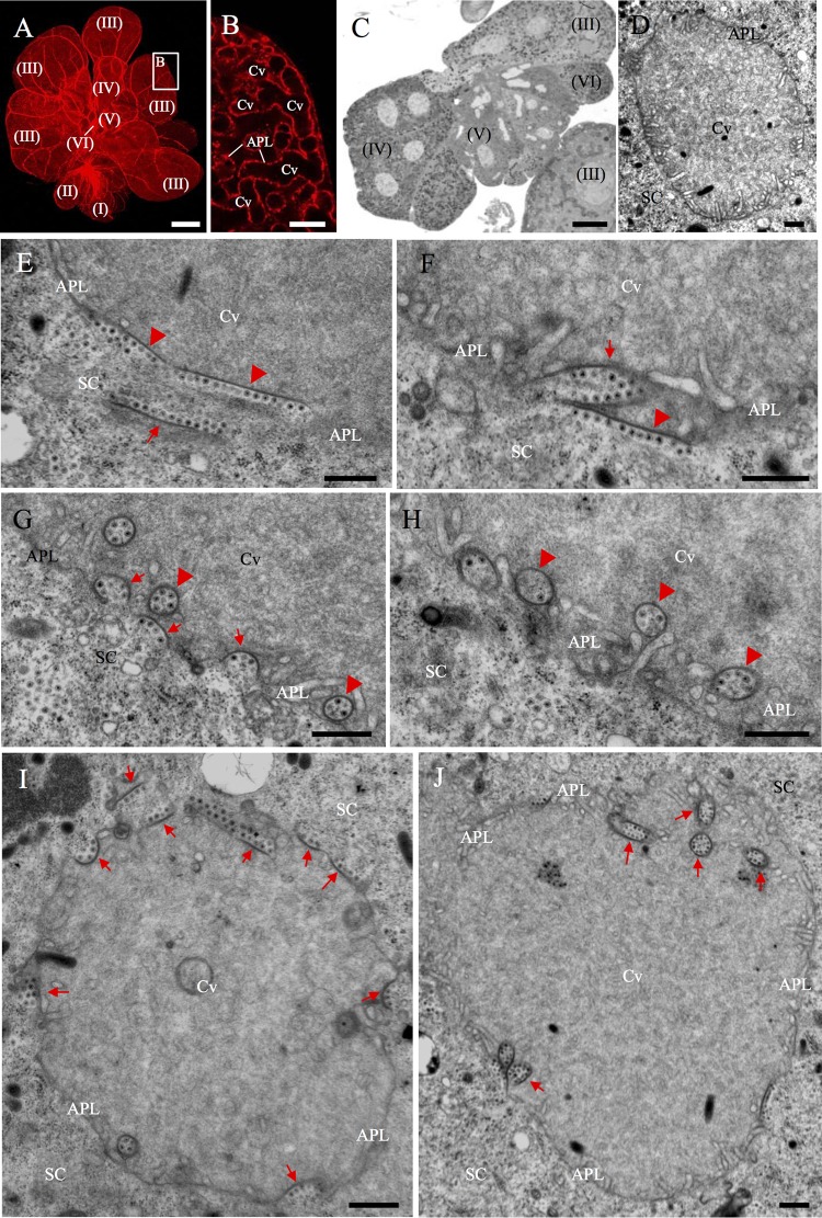
Electron microscopy showing dissemination of RGDV into the apical plasmalemma-lined cavities in the PSGs of R. dorsalis. (A) View of dissected PSG of R. dorsalis, which was stained for actin with phalloidin-rhodamine (red) and then examined via confocal microscopy. The PSG contained six types of secretory cells termed numerically from I-cells (I) to VI-cells (VI). Bar, 100 μm. (B) Enlarged image of the boxed area in panel A, showing the apical plasmalemma-lined cavities forming a loose network within the III-cell. Bar, 10 μm. (C) Semithin section of PSG, showing the cytological characteristics of several types of cells. Bar, 25 μm. (D) Transmission electron micrograph showing the salivary cavity in III-cells. Bar, 500 nm. (E to H) Transmission electron micrographs showing the dissemination of RGDV particles from the cytoplasm of PSG cells to the salivary cavities. RGDV particles first were associated with one edge of filament (arrow) in cell cytoplasm (E). Such virus-associated filaments were attached to the surface of apical plasmalemma (arrowheads; E and F), which induced the formation of invaginations along the apical plasmalemma (arrows; F and G), and finally the formation of vesicular compartments in the cavity (arrowheads; G and H). (I, J) Two virus-associated salivary cavities, showing the presence of virus-associated inclusions (arrows) in the cytoplasm, along the apical plasmalemma, or in the cavity. Bars, 500 nm. APL, apical plasmalemma; Cv, cavity; SC, salivary cytoplasm.

