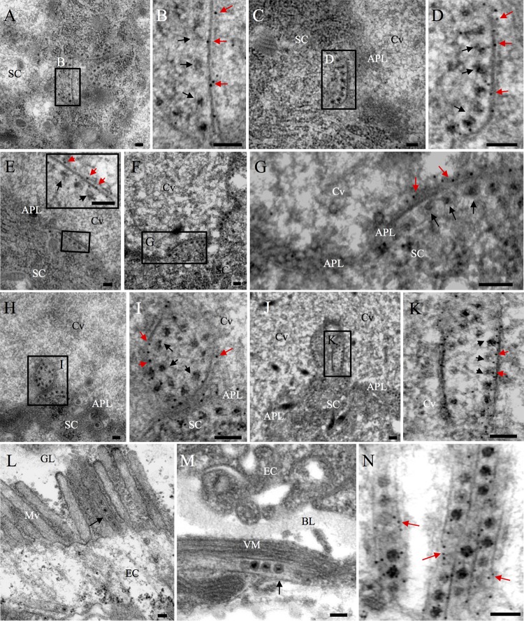
FIG 2.
Immunoelectron microscopy showing the distribution of Pns11 of RGDV during viral infection in the PSGs or midguts of R. dorsalis. (A to K) Immunogold labeling of Pns11 with virus-associated filaments in the cell cytoplasm (A to D), the edges of invaginations along the apical plasmalemma (E to G), or the surface of vesicular compartments in the cavity (H to K). Virus-infected PSGs were immunolabeled with Pns11-specific IgG as primary antibodies, followed by treatment with 10-nm gold particle-conjugated goat antibodies against rabbit IgG as secondary antibodies. Panels B, D, G, I, and K are enlargements of the boxed areas in panels A, C, F, H, and I, respectively. The inset in panel E is an enlarged image of the boxed area in the same panel. Black arrows mark viral particles, while red arrows mark gold particles. (L, M) Association of virus-containing tubules (black arrows) with midgut microvilli (L) or visceral muscles (M). (N) Immunogold labeling of Pns11 with virus-containing tubules in the midgut epithelium. Red arrows mark gold particles. APL, apical plasmalemma; Cv, cavity; SC, salivary cytoplasm; EC, epithelial cells; GL, gut lumen; BL, basal lamina; VM, visceral muscle. Bars, 100 nm.

