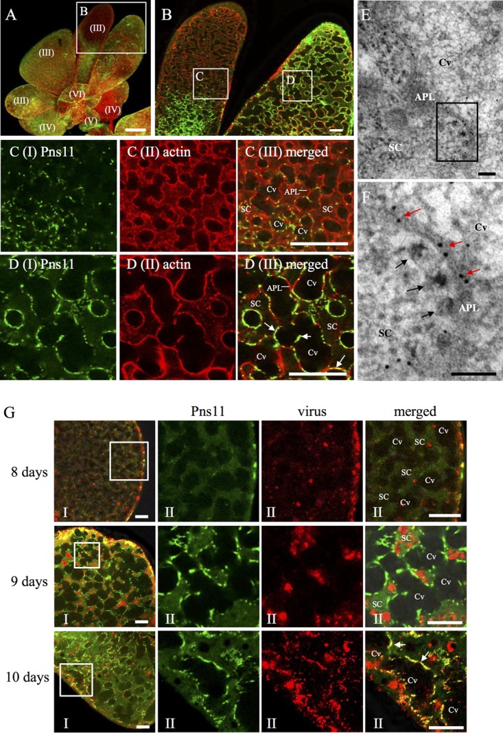
FIG 3.

Immunofluorescence microscopy showing dissemination of RGDV into salivary cavities in the PSGs of R. dorsalis. (A to D) Pns11 of RGDV was closely associated with actin-based cavity plasmalemma in virus-infected PSG regions. PSGs were immunostained with Pns11-FITC (green) and actin dye phalloidin-rhodamine (red) and examined by immunofluorescence microscopy. Panel B shows an enlargement of the boxed area in panel A. Panels C and D show enlargements of the boxed areas labeled C and D, respectively, in panel B. Bar in panel A, 150 μm. Bars in panels B to D, 10 μm. (E, F) Immunoelectron micrographs showing the association of actin with the edges of invagination along the apical plasmalemma. PSGs were immunolabeled with actin-specific IgG as primary antibodies, followed by treatment with 10-nm gold particle-conjugated goat antibodies against mice IgG as secondary antibodies. Red arrows mark gold particles. Black arrows mark viral particles. Panel F shows an enlargement of the boxed area in panel E. Bars, 100 nm. (G) Distribution of RGDV and Pns11 in the secretory cells of PSGs. PSGs at 8, 9, and 10 days padp were dissected and immunostained with Pns11-FITC (green) and virus-rhodamine (red). Panels II show enlargements of the boxed areas in panels I. The arrows mark the association of Pns11 with cavity. Bars, 20 μm. APL, apical plasmalemma; Cv, cavity; SC, salivary cytoplasm.
