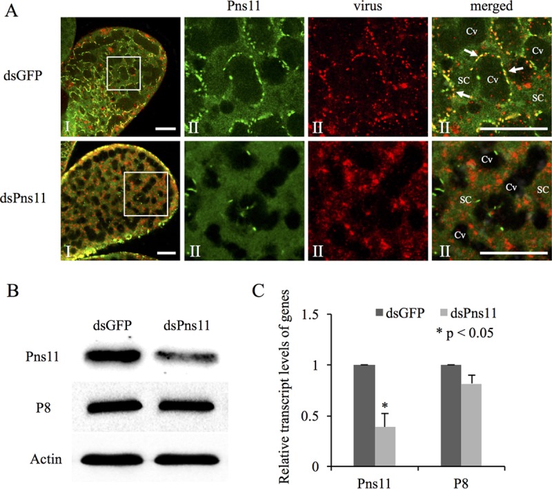
FIG 5.

Microinjection of dsPns11 inhibited Pns11 filament formation and the subsequent transmission of RGDV by R. dorsalis. (A) Three days after microinjection with dsGFP or dsPns11, the PSGs of R. dorsalis were immunolabeled with Pns11-FITC (green) and virus-rhodamine (red) and then examined via confocal microscopy. Panels II show enlargements of the boxed areas in panels I. Cv, cavity; SC, salivary cytoplasm. Bars, 20 μm. All immunofluorescence figures are representative of at least three repetitions. (B) Detection of viral proteins Pns11 and P8 of RGDV in R. dorsalis by Western blotting assay with Pns11-specific or P8-specific IgGs. Insect actin was detected with actin-specific IgG as a control. (C) Detection of transcript levels for Pns11 or P8 genes in R. dorsalis by RT-qPCR assay, with means (±SD) from three independent experiments. *, P < 0.05.
