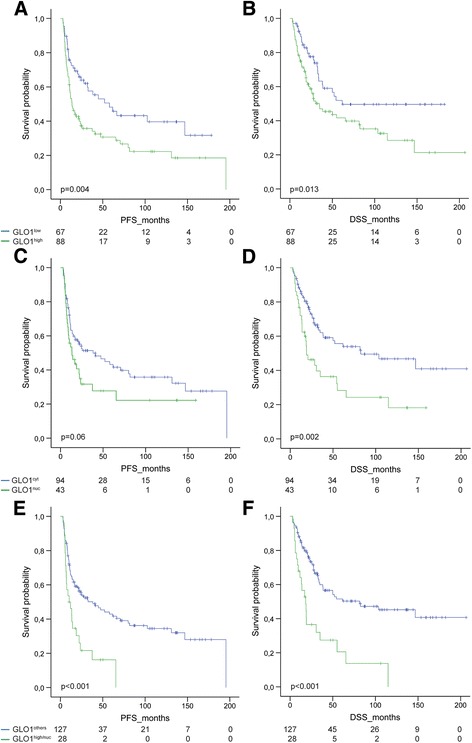
Fig. 3.

High expression and nuclear localization of GLO1 correlates with unfavorable survival. The prognostic value of high versus low GLO1 expression (a-b) and its predominant cytoplasmic versus nuclear localization (c-d) was assessed for progression-free (PFS, left panel) and disease-specific survival (DSS, right panel) in a Kaplan-Meier plot. The worst outcome was observed for OPSCC patients with a combined high and nuclear GLO1 staining as compared to all other staining patterns (e-f). P values were calculated by a log-rank test
