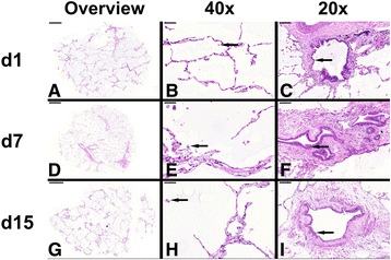
Fig. 5.

Histopathological staining of long-term cultured human PCLS. Representative pictures of human PCLS stained at days 1, 7 and 15 post preparation. The pictures show an overview of PCLS at different time points (a, d & g; left row), close-up pictures of alveolar regions with alveolar macrophages (b, e & h; arrow; middle row, 40× magnification) and close-up pictures of larger airways with bronchiolar epithelium (c, f & i; arrows; right row, 20× magnification)
