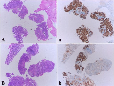
Fig. 1.

Examples of Intratumoral homogeneity and heterogeneity of HER2 IHC staining in biopsy specimens. A The biopsy of an intestinal type GC shows 4 tumor-containing fragments in the 5 biopsy specimens. a HER2 IHC staining shows that all the 4 tumor fragments are uniformly 3+ (homogeneous). B Four tumor fragments are found in this biopsy of an intestinal type GC. b Within the 4 fragments, 3 are stained 3+, the 4th fragment is stained 1+ (heterogeneous). In addition, within the 3 HER2 3+ fragments, one of them demonstrates focally positive
