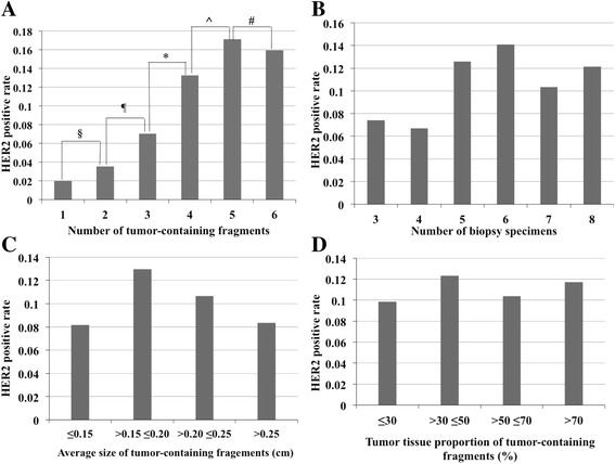
Fig. 2.

The associations of HER2 IHC positive (scored 3+) rate with tumor-containing fragment number, biopsy specimen number, average size and tumor tissue proportion of tumor fragments. a The HER2 IHC positive (scored 3+) rate exhibited elevation with increased tumor fragment numbers (P = 0.004). The rates of 1, 2, 3, 4, 5 and 6 fragments were 2.0, 3.5, 7.0, 13.2, 17.1, and 15.9%. The 4-fragment subgroup showed a much higher HER2 IHC positive rate than the 3-fragment subgroup. No statistical difference is reached among subgroups with 4, 5 and 6 fragments. § P = 0.6, ¶ P = 0.263, *P = 0.035, ^P = 0.299, # P = 0.812. b HER2 IHC positive (scored 3+) rates were 7.4, 6.7, 12.6, 14.1, 10.3, and 12.1% when the biopsy number was 3, 4, 5, 6, 7, and 8 respectively without significant differences (P = 0.127). c HER2 3+ rates of the subgroups divided based on average size of tumor fragments were 8.2, 12.9, 10.7 and 8.3% without statistical difference (P = 0.397). d HER2 3+ rates of subgroups divided based on tumor tissue proportion of tumor fragments were 9.8, 12.3, 10.4 and 11.7% (P = 0.825)
