
Fig. 1.
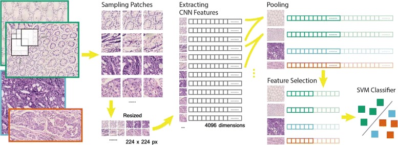
The classification workflow. First, square patches of 336 or 672 pixels in size are sampled on a rectangular grid, depending on the magnification scale of the image. Patches are then resized to 224 pixels in size as the input of our CNN model. A 4096-dimensional feature vector is extracted from the CNN model for each patch. A 100-dimensional feature is obtained by feature pooling and feature selection for each image. Finally, a linear SVM classifies the selected features. The figure shows a binary classification, where the positive (blue and orange) and negative (green) are GBM and LGG in brain tumor, cancer and normal in colon cancer respectively. In multiclass classification, a full feature vector of 4096 dimensions is used