Figure 1.
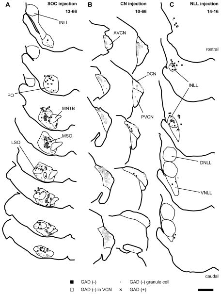
Traces of injection sites of recombinant viruses. Filled squares indicate GFP+ neurons negative for GAD67; “x”, GFP+ neurons positive for GAD67; open squares, GFP+ VCN neurons negative for GAD67; dots, GFP+/GAD67− neurons in the granule cell domain of the CN. (A) An example of SOC injection cases (13-66). Infected neurons were distributed throughout the SOC. The few infected cells outside of the SOC were not in auditory nuclei. (B) An example of CN injection cases (10-66). Infected neurons were found in the DCN, AVCN, and PVCN. (C) An example of INLL injection cases (14-16). Infected neurons were mainly found in the INLL and the surrounding regions. Each trace is separated by 240 μm. Scale bar: 1 mm. Abbreviations: AVCN, anteroventral cochlear nucleus; DCN, dorsal cochlear nucleus; DNLL, dorsal nucleus of the lateral lemniscus; INLL, intermediate nucleus of the lateral lemniscus; LSO, lateral superior olive; MSO, medial superior olive; MNTB, medial nucleus of the trapezoid body; PO, periolivary nucleus; PVCN, posteroventral cochlear nucleus; VNLL, ventral nucleus of the lateral lemniscus.

