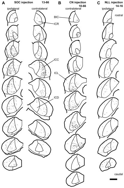
Figure 2.
Distribution of LG cells (dots) which received axosomatic contacts with GFP+/VGLUT2+ terminals. (A) An SOC injection case (13-66). LG cells receiving contact were found bilaterally. In the ICC, LG cells receiving contact were located mainly in the lateral part. (B) A CN injection case (10-66). LG cells receiving contact were mainly found in the ICC of the contralateral side. (C) An NLL injection case (14-16). LG cells receiving contact were sparsely distributed throughout the IC of the ipsilateral side. Each trace is separated by 240 μm. Scale bar: 1 mm. Abbreviations: BIC, brachial nucleus of the IC; ICC, central nucleus of the IC; ICD, dorsal cortex of the IC; ICL, lateral cortex of the IC; ICR, rostral cortex of the IC.

