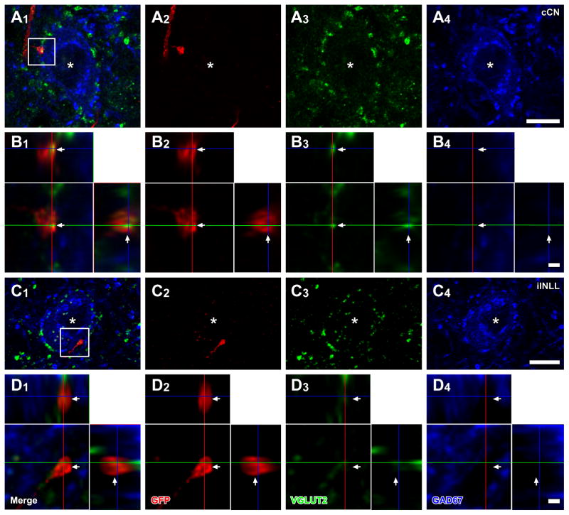
Figure 4.
LG cells receive axosomatic excitatory inputs from the cochlear nuclei and INLL. LG cells (blue, asterisks) with GFP+ (red) terminals, which colocalized VGLUT2 immunoreactivity (green), were found after an injection of palGFP Sindbis virus in the contralateral CN (case 10-68; A, B), or ipsilateral INLL (case 11-22; C, D). (B, D) High-magnification images of a box in A1 and C1 with orthogonal views of the stack cut at 3 planes parallel to xy- (blue lines), yz- (a red line), and xz-planes (a green line). GFP+/VGLUT2+ terminals made contact on LG cells (arrows). Scale bars: 10 μm (A, C), and 1 μm (B, D). Note that three-dimensional reconstructions of these GFP+ axons and LG cell bodies are shown in Fig. 5.

