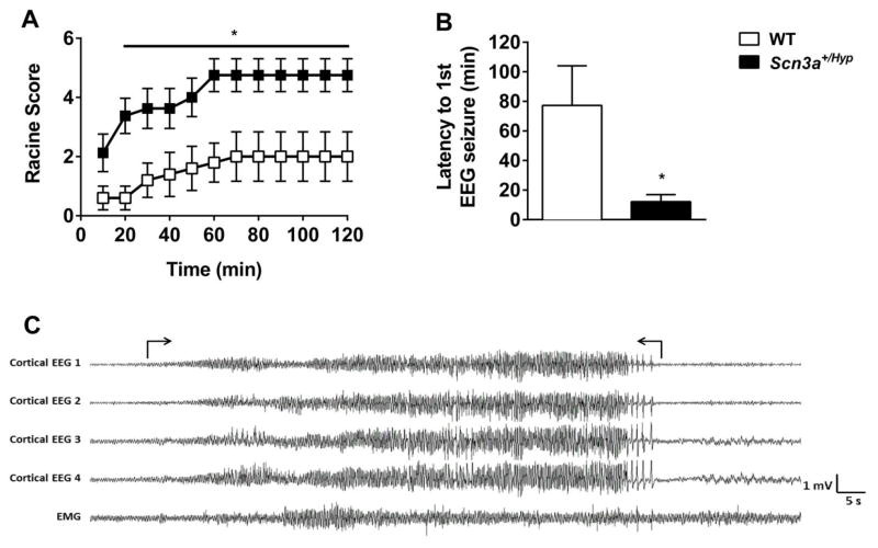
Fig. 7.
Scn3a+/Hyp mice exhibit increased susceptibility to KA-induced seizures. (A) Following the administration of KA, Scn3a+/Hyp mutants (n = 8) exhibited significantly more severe seizure phenotypes when compared to WT littermates (n = 5). *p < 0.05; Two-way repeated measures ANOVA. (B) Average latency to the first electrographic seizure was shorter in Scn3a+/Hyp mutants. *p < 0.05, Two-tailed Student’s t-test. (C) A representative EEG recording during a KA-induced seizure in a mutant mouse. Seizure onset and termination are indicated within brackets. EEG montage: Cortical EEG 1, Cortical EEG 2, Cortical EEG 3, and Cortical EEG 4 – cortical electrodes. EMG – muscle electrodes. All cortical electrodes and EMG referenced to a second EMG electrode. Error bars indicate SEM.

