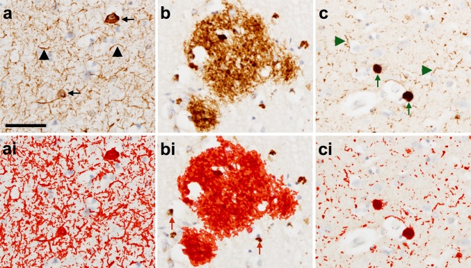
Fig. 3.
Photomicrographs illustrating immunohistochemically stained pathology (a–c) and the application of a standardized threshold designed to capture all immunopositive signals to be included in the quantitative analysis (red outline in ai–ci). Neurofibrillary tangles (black arrow) and neuropil threads (black arrowhead) are immunopositive for HP-T (AT8 antibody) (a) and with threshold applied—red outline (ai). Plaques are immunopositive for β amyloid (4G8 antibody) (b) and with the threshold applied—red outline (bi). Intracellular amyloid precursor protein is also immunopositive using 4G8 antibody and is excluded from the quantitative analysis using a size restriction threshold (red arrows) (bi). Lewy bodies (green arrows) and Lewy neurites (green arrowheads) are immunopositive for α-syn (α-syn antibody) (c) and with threshold applied—red outline (ci). Scale bar in a represents 50 μm and is valid for all images

