
Fig. 1.
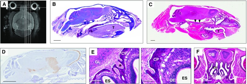
Whole head mounting allows for visualization of the intact olfactory system by routine microscopy. a Magnetic resonance image of an adult mouse head with sagittal and coronal lines (dotted) depicting examples of levels for sectioning. b Luxol fast blue-stained, and c haemotoxylin and eosin (H&E)-stained, sagittal head sections (all, 5 μm) from an adult mouse that visualize, among other structures: the brain with an intact olfactory bulb (b more medial; c lateral), turbine within the ethmoid sinus, the palate, tongue (“T”), alveolar bone (and teeth), an intact olfactory epithelium including cranial nerve (CN)-I fibers as well as CN-V at the base of the skull (c arrows). d Tyrosine hydroxylase immunohistochemistry-based staining of an intact skull section from an adult mouse (at an intermediate level) highlighting the Substantia nigra of the midbrain, neostriatum, and dopamine-producing neurons of olfactory glomeruli (brown staining). e Higher magnification of H&E stained, sagittal section of the olfactory system in an adult mouse. ES ethmoidal sinus, CP cribriform plate, GL glomeruli; asterisk denotes axonal bundles of CN-I. f H&E staining of an adult mouse skull prepared for coronal sectioning at the level of the olfactory bulb (OB) and turbine/ES, as depicted in a. Scale bars represent 2 mm (b–d, f); 200 μm (e, left image); 100 μm (e, right image)