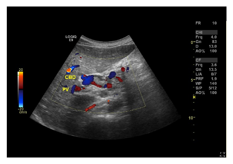
Figure 1.

Abdominal ultrasound (post-ERCP with stenting) showing multiple intraluminal stones in the gallbladder with thick biliary sludge and diffusely thickened wall. Biliary stent is seen with the visualized portion of distal CBD that also appears slightly dilated at the porta hepatis. Minimal central intrahepatic biliary dilatation is also seen. Status after splenectomy.
