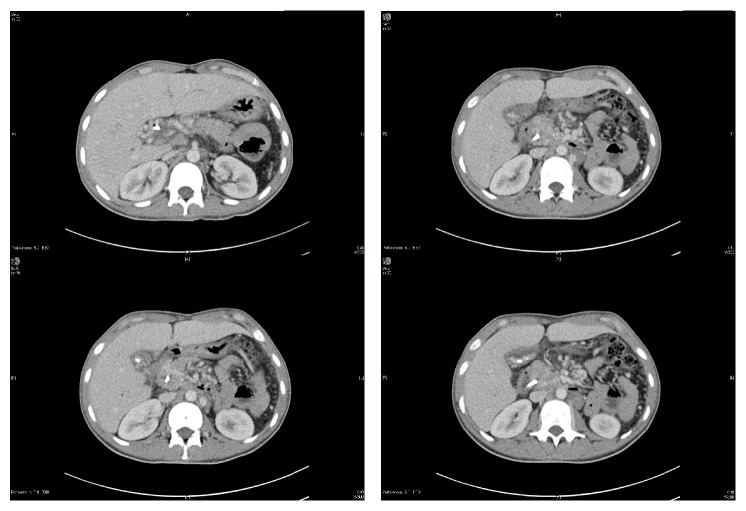
Figure 2.
Multiple abdomen computed tomography (CT) scans showing the development of multiple collaterals in the porta hepatis replacing the portal vein, suggestive of cavernous transformation due to chronic portal vein thrombosis, in addition to multiple mesenteric and upper retroperitoneal collaterals. Multiple gallbladder stones and CBD stent are also seen. The liver is seen slightly enlarged with no detectable focal lesions. Normal enhancement of the hepatic veins. Normal size and parenchymal density of the pancreas. No detected gross masses. No detected calcification. Clear peripancreatic fat planes. Status after splenectomy.

