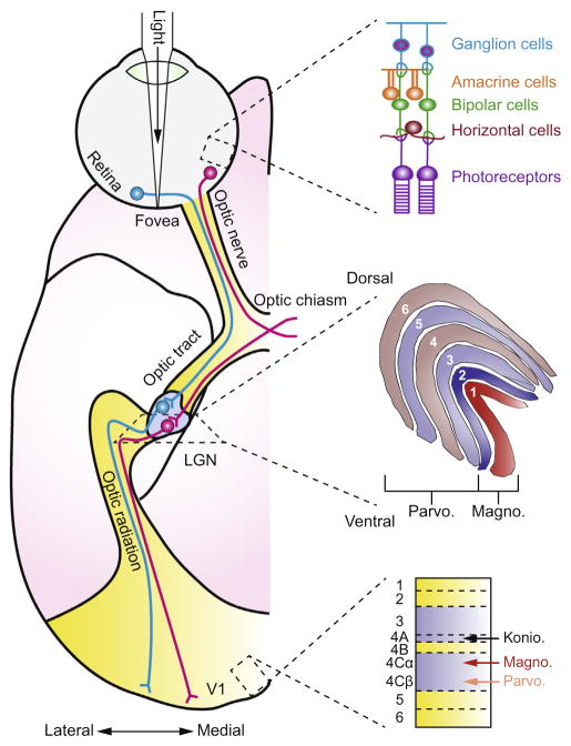
Fig. 1.
Early visual system pathways of the macaque monkey. The figure on the left shows the pathway of visual information imaged on the retina as it passes through the LGN and arrives at the primary visual cortex (V1). The anatomical schematic represents a ventral view of the right hemisphere. The visual scene is imaged by photoreceptors in the retina and information is passed through bipolar cells to retinal ganglion cells whose axons exit the back of the eyeball forming the optic nerve. Information from the contralateral part of the scene reaches the LGN with input from the two eyes arriving at separate layers of the LGN: layers 2, 3, and 5 receive input from the ipsilateral eye and layers 1, 4, and 6 receive input from the contralateral eye. The magnocellular layers (1 and 2) receive input that originated from rod photoreceptors and the Parvocellular layers (3–6) receive input that originated from cone photoreceptors. Koniocellular cells in the LGN are interspersed between the magnocellular and parvocellular layers and receive information arising from short-wavelength cones. Cells in the LGN project mainly to layer 4 of the primary visual cortex through a formation called the optic radiation. Adapted from Solomon and Lennie, 2007 with permission.

