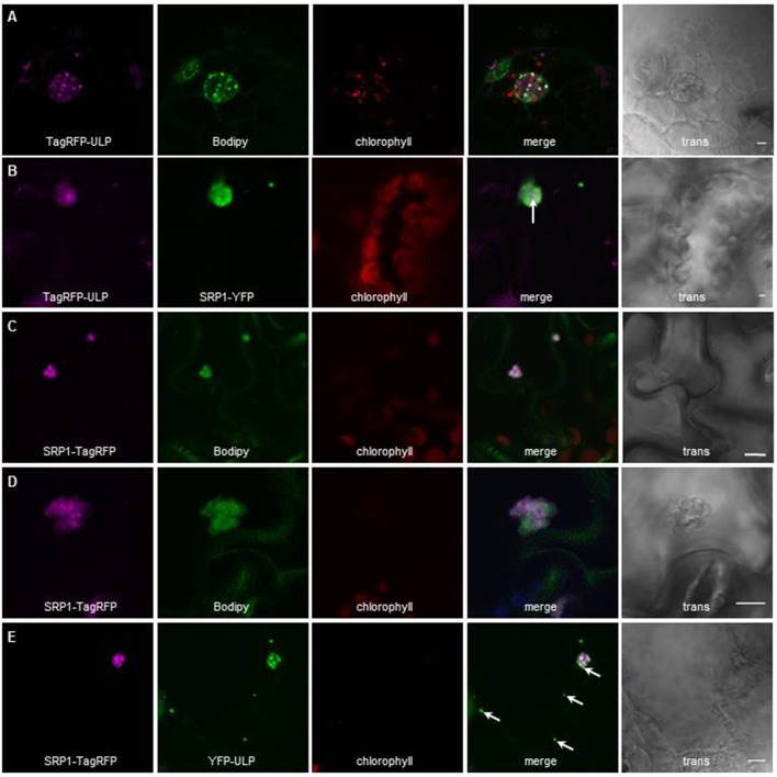
Figure 2.
Localization of AtULP and AtSRP1-fluorescent fusions in lipid droplets. Arabidopsis cotyledons (A,C–E), or N. benthamiana leaves (B) expressing TagRFP-AtULP (A,B) AtSRP1-YFP (B), AtSRP1-TagRFP (C–E), or YFP-AtULP (E) were visualized by confocal laser scanning microscopy. Arabidopsis plantlets were co-labeled with neutral lipid specific dye BODIPY493/503(A,C,D). Co-labeling of AtULP or AtSRP1 targeted structures by BODIPY493/503 confirmed that these structures are lipid droplets. Bodipy, chlorophyll, and trans indicate Bodipy fluorescence, chlorophyll autofluorescence and transmission microscopy image respectively. Merge indicates merge of TagRFP, Bodipy and chlorophyll fluorescences (A,C,D) or YFP and TagRFP fluorescence (B,E). Bar: 5 μm. White arrows indicate specific localization of AtULP in cells expressing both AtSRP1 and AtULP transgenes.

