Figure 6.
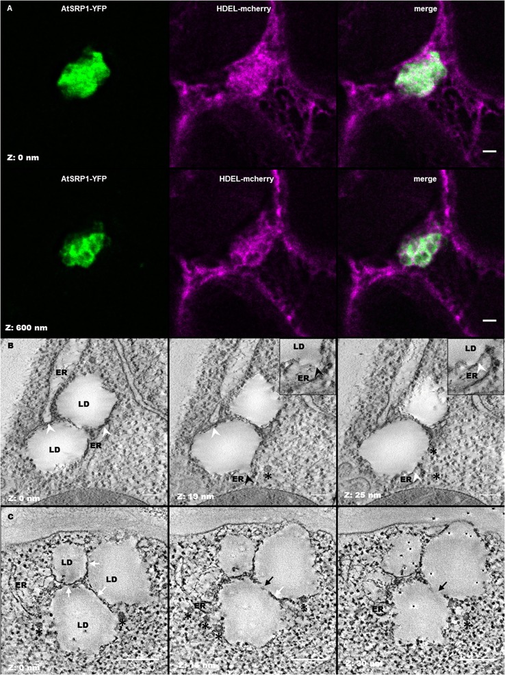
Leaf lipid droplets (LDs) are in close proximity with the endoplasmic reticulum (ER). (A) Fluorescent co-labeling of leaf LDs and the ER. N. benthamiana leaves were transiently co-transformed with AtSRP1-YFP and HDEL-mCherry constructs labeling LDs and ER membranes respectively. Deconvolution after confocal imaging of epidermal cells demonstrates multiple contact sites between LDs and the ER network, and AtSRP1-YFP localization at the LD periphery. Merge indicates overlap of YFP (green) and mCherry (magenta) fluorescences. Bar: 2 μm. (B,C) Electron tomography of leaf LDs in AtSRP1 overexpressing Arabidopsis plantlets with single (B) and dual (C) axis acquisition. White arrowheads indicate LD/ER contacts. Black arrowhead indicates the ER mono-leaflet at the LD budding point. White arrows indicate apposition of two LDs, black arrows indicate direct continuity between two LDs. Black asterisks are positioned below vesicle-like structures. Black points with white halo in last panel are gold fiducials used for alignment steps during tomogram. Bar: 200 nm.

