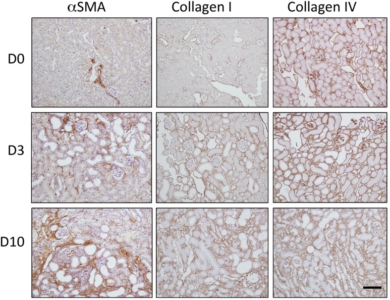
FIGURE 1.
Progression of fibrosis in the mouse after unilateral ureteric obstruction (UUO). Immunohistochemical staining for αSMA (a marker of myofibroblast recruitment) and the extracellular matrix proteins collagen I and collagen IV. Representative micrographs are shown from normal kidneys (D0) and kidneys 3 days (D3) and 10 days (D10) post-UUO. Nuclei were counterstained with hematoxylin. Scale bar = 100 μm.

