Figure 1.
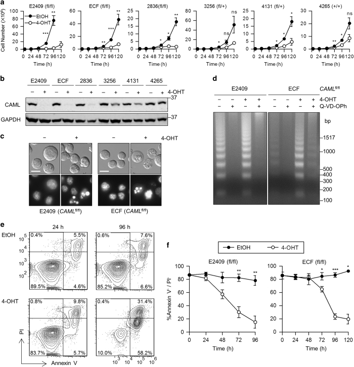
Caml-deleted Eμ-Myc lymphoma cell lines display impaired proliferation and apoptotic hallmarks. (a) Growth curves for Eμ-Myc;Cre-ER;Camlfl/fl, Camlfl/+, or Caml+/+ lymphoma cell lines treated with vehicle (EtOH) or 4-OHT. Live cells were counted at 24 h intervals by flow cytometry. (b) Loss of CAML protein in 4-OHT-treated Eμ-Myc;Cre-ER;Caml-floxed lymphoma cell lines. Lysates were collected from parallel cultures of a at 48 h, and CAML expression was analyzed by western blot. (c) Apoptotic nuclear condensation observed by Hoechst 33342 staining of 4-OHT-treated E2409 and ECF cells. Scale bar=10 μm (d) DNA laddering in apoptotic 4-OHT-treated cells, with rescue by pan-caspase inhibitor treatment (Q-VD-OPh, 5 μM). (e) Staining with annexin V and propidium iodide (PI) indicate apoptotic cell death in Caml-deleted cells. Eμ-Myc;Cre-ER;Camlfl/fl (E2409) cell line was treated with vehicle (EtOH) or 4-OHT, and samples were stained at 24 and 96 h with annexin V-Cy5 and PI. (f) Live cells (annexin V-negative and PI-negative) declined over time following 4-OHT treatment of Eμ-Myc;Cre-ER;Camlfl/fl cell lines compared with vehicle control. Values are mean±S.E.M. of three independent replicates. Statistical differences between EtOH and 4-OHT treatments, for each time point starting at 72 h, are denoted by ‘ns’ for not significant, *P<0.05, **P<0.01, and ***P<0.001.

