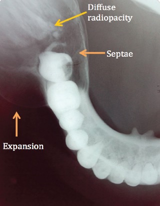
Figure 3.

Mandibular occlusal radiograph reveals a well-defined expansion of both the buccal and lingual cortical plates arising from lower right 1st molar region, with evidence of septa suggesting a multi-locular appearance and diffuse irregular radiopacity within the largely radiolucent lesion
