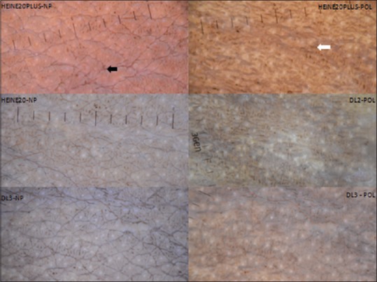
Figure 1.

Dermatoscopy of Acanthosis nigricans over the neck (10×) – Non-polarized images show multiple cristae and sulci clearly (black arrow) whereas polarized imaging allows better visualization of hyperpigmented dots and streaks (white arrow). Polarized images taken with Heine Delta 20 plus have a warmer tone whereas DL3 polarized images are the brightest
