Figure 3.

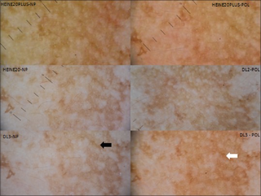
Dermatoscopy of Melasma over the face (10×) – Polarized images show the accentuation of pseudoreticular pattern, which is more brownish (white arrow) than nonpolarized images (black arrow). Heine delta 20 plus images are warmer

Dermatoscopy of Melasma over the face (10×) – Polarized images show the accentuation of pseudoreticular pattern, which is more brownish (white arrow) than nonpolarized images (black arrow). Heine delta 20 plus images are warmer