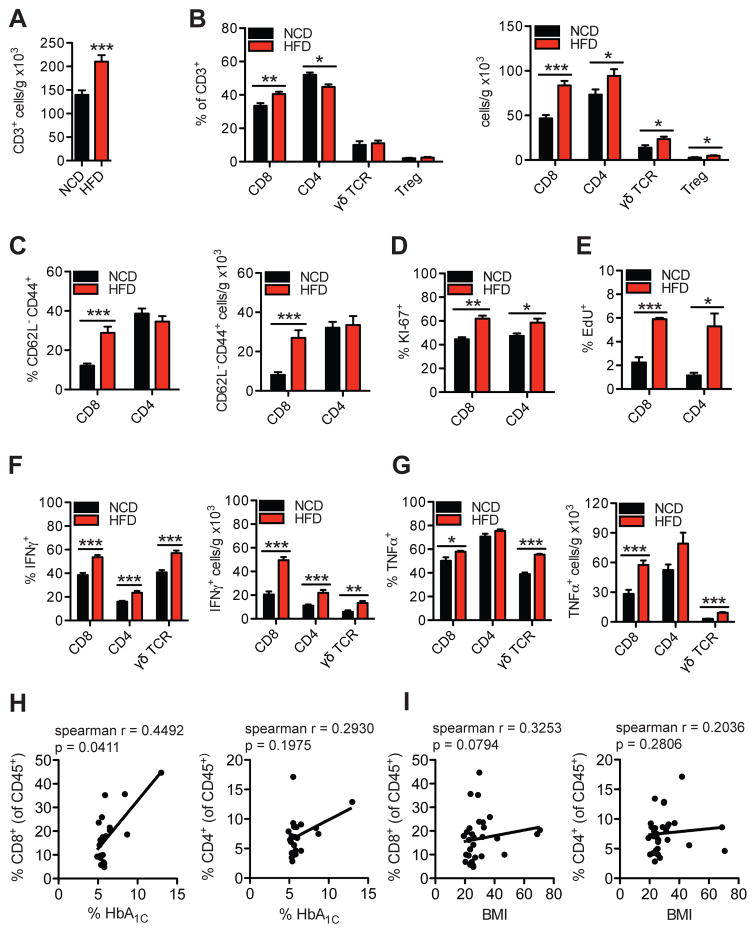
Figure 1. HFD-feeding promotes a pro-inflammatory shift in hepatic T cell populations in mice and hepatic CD8+ T cells correlate to glucose dysregulation in humans.
Flow cytometry analysis of liver immune cells isolated from 16 wk HFD- or NCD-fed WT mice representing (A) total number of hepatic CD45+CD3+NK1.1- gated T cells per gram of tissue (n=16), (B) CD8+, CD4+, γδTCR+, or CD4+Foxp3+ (Treg) gated hepatic T cell subsets (n=5–16), (C) CD8+ or CD4+ CD62L− CD44+ gated effector memory T cell subsets (n=15), (D–G) intracellular T cell stains for (D) Ki-67 (n=4), (E) EdU (n=4), (F) IFNγ (n=9), and (G) TNFα (n=4) in CD8+, CD4+ or γδTCR+ gated T cells. Correlative analysis between (H) % CD8+ and % HbA1c (left), % CD4+ and HbA1c (right), and (I) % CD8+ and BMI (left), % CD4+ and BMI (right) from human intrahepatic mononuclear cells (HbA1c n=21, BMI n=30). Spearman r values and p values are denoting statistical significance are indicated in the figure graphs. Statistical significance is denoted by * (p<0.05), ** (p<0.01), and *** (p<0.001).

