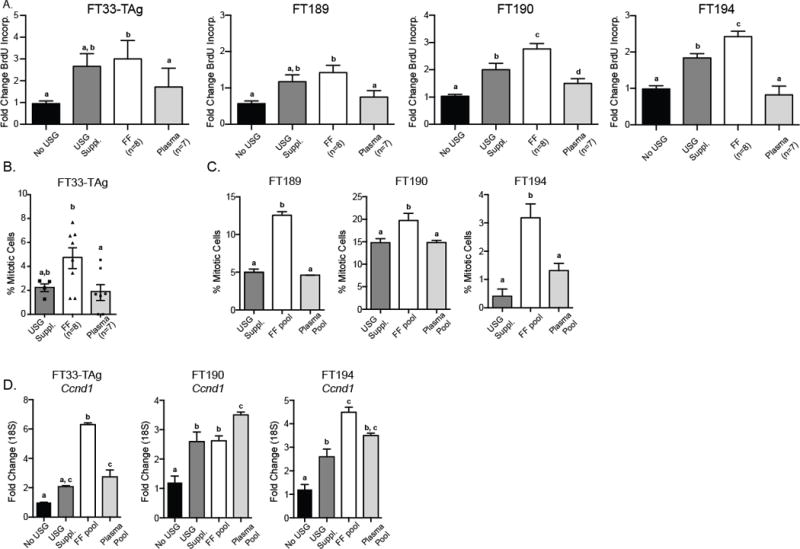
Figure 1. Fallopian epithelial cell proliferation following follicular fluid or plasma exposure.

A) Cell lines were cultured in basal medium (No USG), USG supplement (USG Suppl.), 5% individual follicular fluid, or corresponding patient plasma (5%) for 24 hr and BrdU incorporation was measured. B) Microscopy image analysis of mitotic figures performed on FT33-TAg cells exposed to individual patient FF and matched plasma samples. C) Percentage of mitotic cells in the FT189, FT190 and FT194 cells grown in USG Suppl., pooled FF, or pooled plasma from all 8 patients (three independent assays). Mitotic figures were counted based on chromosome Hoescht staining. D) Proliferative index as assessed by Cyclin D1 (Ccnd1) expression following growth of FT33-TAg, FT190 and FT194 cells for 24 hr in basal medium (No USG), USG Suppl., FF pool, or matching patient plasma pool. a,b,c Means ± SEM within a panel that have different superscripts were different (p<0.05).
