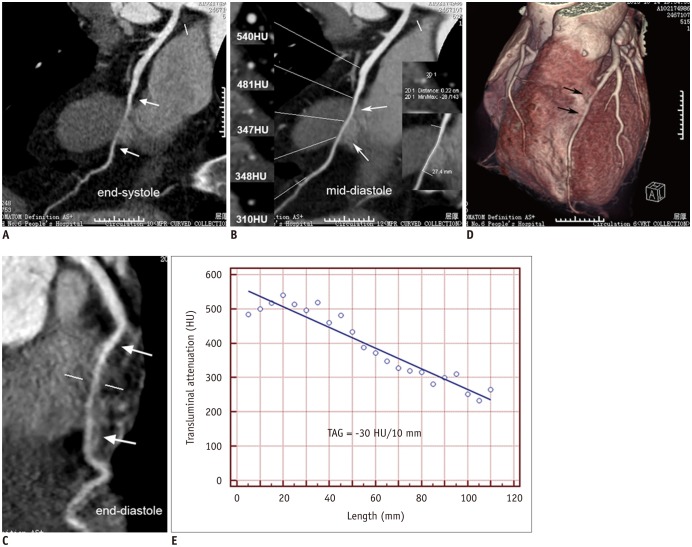
Fig. 2. Representative case of MB with dynamic compression.
A. CPR image of end-systolic phase (35% of R-R interval) showed presence of MB at middle LAD with significant compression (white arrows). B. CPR image of mid-diastolic phase (70% of R-R interval) showed presence of MB at middle LAD (white arrows). MB depth and length were 2.2 and 27.4 mm, respectively. C. CPR image of end-diastolic phase (5% of R-R interval) showed presence of MB at middle LAD (white arrows) with persistent compression. D. VR image confirmed overlay of myocardium at middle LAD (black arrows). E. TAG of MB vessel was -30 HU/10 mm. CPR = curved planar reformation, HU = Hounsfield units, LAD = left anterior descending, MB = myocardial bridge, TAG = transluminal attenuation gradient, VR = volume rendering

