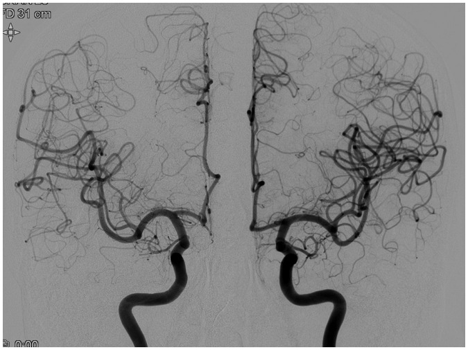
Fig. 1. Anteroposterior views of bilateral paired internal carotid artery (ICA) injections (Rt ICA, protocol 4; Lt ICA, protocol 1).
Number of exposures, field of view, table height, source to distance, and tube angulations were matched for both injections. Despite lack of perceptible difference in image quality, about 40–50% of total AK and 25–40% of total DAP reduction was seen in patients studied with protocol 4. AK = air kerma, DAP = dose area product

