
Figure 7.
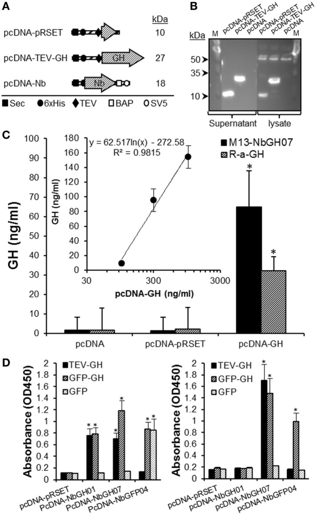
Quantitation of GH in the supernatant of HEK293 cell using M13-NbGH07 phage enzyme-linked immunosorbant assay (ELISA). (A) Schematic representation of the recombinant proteins; pRSET, TEV-GH, and nanobodies secreted by HEK293 transfected cells with the corresponding pcDNA plasmids. The theoretical molecular size (kiloDaltons) is shown to the right of each recombinant construct. Positions of the different elements, the Sec signal, 6 × His tag, TEV, BAP, and SV5, are indicated using specific symbols ■, ●, ♦, □, and ○, respectively. (B) Detection of pRSET and TEV-GH proteins in the lysate (10 µl) and supernatant (25 µl) of 48 h transfected HEK293 cells after SDS-PAGE (15%) separation by immunoblotting using anti-6 × His antibody. (C) Measuring of GH (nanograms per milliliter) in the cell supernatants was performed using phage (model IV) or conventional (model III) sandwich ELISA. (Inset) Quantitation of GH concentration (nanograms per milliliter), using phage ELISA (model III), in the supernatant (25 µl) of HEK293 cells after 48 h of transfection with serial concentrations of pcDNA-GH plasmid. (D) Biotinylated nanobodies (NbGH01, NbGH07, and NbGFP04) secreted in the supernatant of transfected cells were tested in conventional (model I where 6 × His tag is replaced by the biotin group, left panel) and sandwich (model IV where M13 is replaced by the biotin group and using NbGH04 as bait, right panel) ELISA. Streptavidin–HRP (1:1,000) was used to reveal the biotinylated detector nanobodies bound to GH, GFP-GH, and GFP antigens.