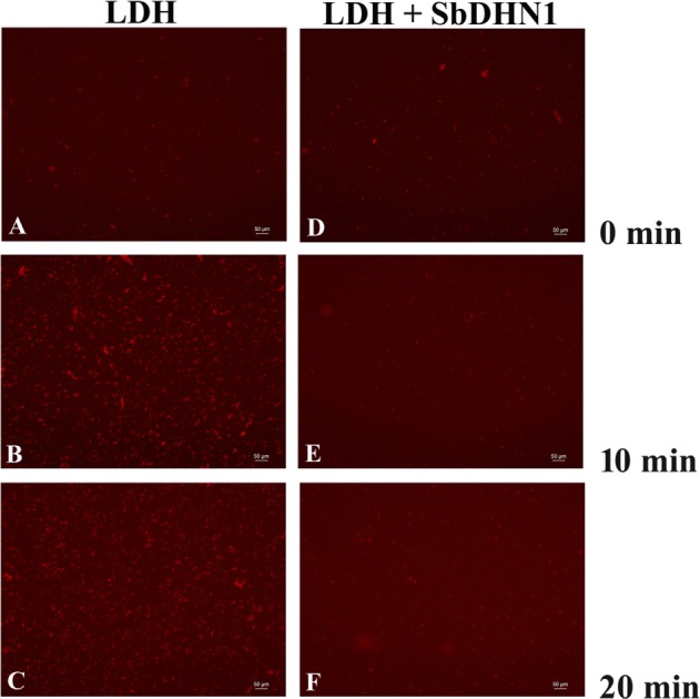
FIGURE 8.

Lactate dehydrogenase was subjected to high temperature (60°C) in presence and absence of SbDHN1 protein in a time dependent manner (0, 10, and 20 min). Fluorescence microscope images were obtained after staining with Congo Red solution. (A–C) LDH in absence of SbDHN1 protein at 0, 10, and 20 min, respectively. (D–F) LDH in presence of SbDHN1 protein at 0, 10, and 20 min, respectively.
