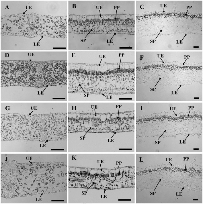
Figure 3.
Leaf sectioning anatomy of C. australis (left panel), F. benjamina (middle panel), and S. speciosa (right panel) developed under Red light (A–C), Blue light (D–F), Red with Blue (G–I) and White (J–L). Black bar = 100 μm. UE, upper epidermis; LE, lower epidermis; PP, palisade parenchyma; SP, spongy parechyma.

