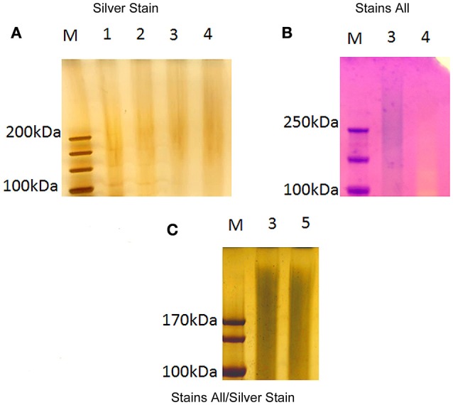
Figure 2.

Electrophoretic profile of large molecular size F. novicida extracts. Extracts from F. novicida were separated by electrophoresis through a 4–12% SDS-PAGE gel. The gels were subsequently stained by silver stain (A), Stains-All (B), and Stains-All/silver (C). LVS CLC was used for comparison. Analysis of the profiles focused on material >100 kDa. Lanes: M, molecular size standards; 1, LVS crude CLC in 1 M urea; 2, F. novicida crude extract in 1 M urea; 3 F. novicida soluble fraction extract (urea extracted); 4, F. novicida insoluble fraction extract (urea extracted); 5, F. novicida soluble fraction extract (phenol extracted).
