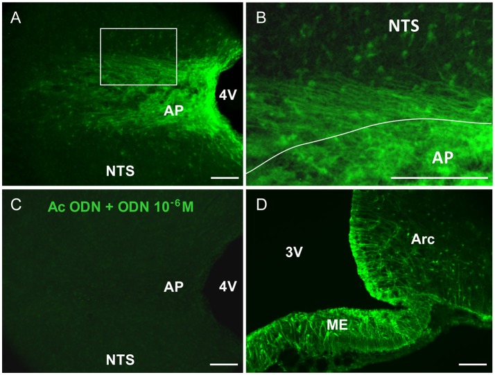
Figure 1.
ODN immunohistochemistry in the brainstem. (A) Confocal fluorescent micrographs illustrating ODN immunohistochemistry performed on horizontal brainstem sections. ODN immunostaining showed the presence of positive cells in the area postrema (AP), at the border between AP and nucleus tractus solitarius (NTS) and within NTS surrounding the AP. Lateral dorsal vagal complex was devoid of labeling. (B) High magnification microphotograph originating from image in A (rectangle in A) and illustrating the shape of ODN immunoreactivity within the AP and surrounding structures. (C) Pre-incubation of ODN antibody with an excess of ODN peptide resulted in the absence of staining. (D) Confocal fluorescent micrograph illustrating ODN immunohistochemistry performed on coronal hypothalamic sections. As expected ODN immunoreactivity was found in the median eminence (ME), tanycytes lining the 3th ventricle (3V) and arcuate nucleus (Arc). 4V, 4th ventricle. Scale bars: 100 μm.

