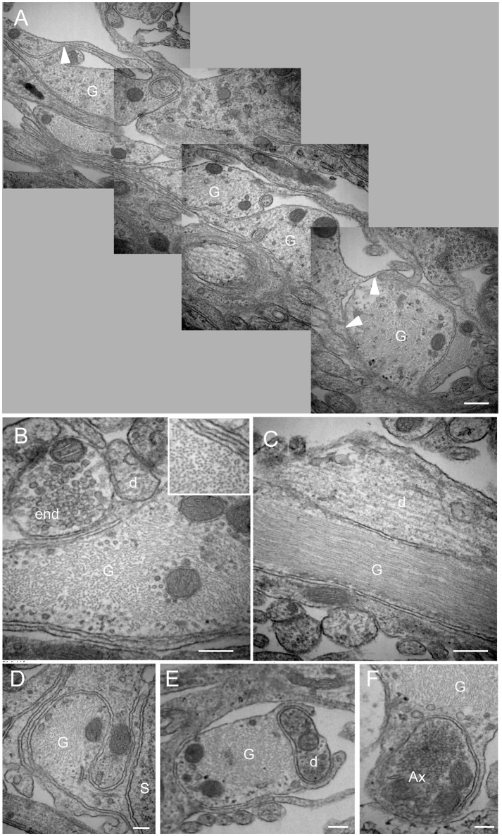
Figure 4.
Morphological organization of the NTS/AP border. (A) Electron micrographs performed on sagital dorsal vagal complex sections showing the presence of rounded, fibrous glial processes at the border between the area postrema (AP) and nucleus tractus solitarius (NTS). Note that these fibrous processes create a continuous layer between the AP and the NTS. Small ramifications (arrowheads) of the rounded processes could be observed. (B,C) These glial processes could be identified by their numerous cytoskeleton filaments visible on sagital (B) and horizontal (C) sections. Inset in (B) Illustration of the high cytoskeleton filaments density in rounded glial processes. (D–F) Close juxtapositions between fibrous glial processes and neuronal elements i.e., neuronal soma (D), dendritic profile (E) and axon terminal (F) are noticeable. Scale bars: 500 μm in A; 200 μm in (B–F). D, dendritic profiles; Ax, axon terminals; S, soma; G: glial process.

