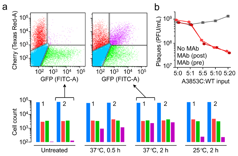
Fig. 2. Functional implications of multi-virion infectious units.
Proof of concept for the co-transmission of genetic variants was obtained by flow cytometry using two fluorescently labelled viruses (a). Cells were inoculated with VSV-mCherry and VSV-GFP, incubated 7 h, and counted (105 events per assay). Bottom: uninfected (blue), mCherry-positive (red), GFP-positive (green), and doubly fluorescent (purple) cell counts following direct inoculation with VSV-GFP and VSV-mCherry (untreated) or after co-incubation of the two virion types (0.5 h at 37°C, 2 h at 37°C, or 2 h at 25°C). Two independent assays (1, 2) are shown for each condition. Top: flow cytometry scatter plot from two of the experiments. The estimated fraction of total virions forming dual infectious units was inferred using a probabilistic model detailed in Supplementary Fig. 3. Data are provided in Supplementary Table 1. b. Effects of virion-virion binding on antibody escape. Plaque assays of the MAb-resistant (A3853C) and MAb-sensitive (WT) variants following pre-incubation of the two types of virions (37°C, 2 h) at different proportions (v:v): 5:0, 5:1, 5:5, 5:10, and 5:20. For each treatment, plaque assays were done in the absence of MAb (grey), adding MAb to the culture medium to inhibit plaque development (dark red) or adding MAb prior to cell inoculation to inhibit cell entry (light red). Data points indicate average titers (PFU/mL) obtained from three independent assays, and error bars indicate the standard error of the mean.

