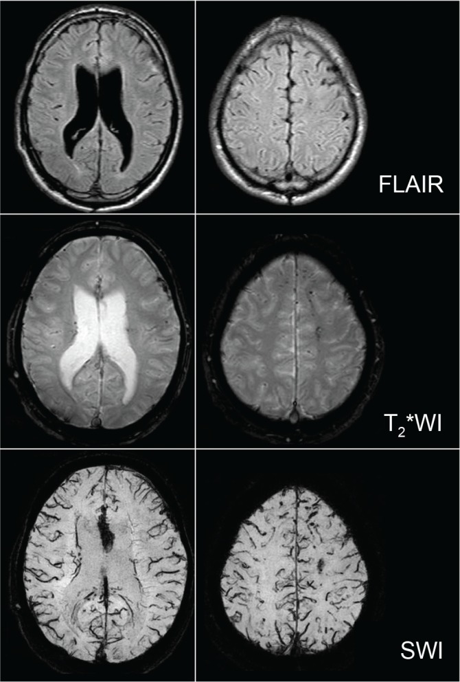
Fig. 3.

MRI of a patient with severe diffuse brain injury in the chronic phase. Hemosiderin can be well visualized on T2* weighted imaging (T2*WI) and susceptibility weighted imaging (SWI) compared to fluid attenuated inversion recovery (FLAIR) imaging. Especially, hemosiderin in the corpus callosum and cerebral white matter can be more conspicuously seen on SWI.
